We tend to file energy and hormones in separate departments. One is about feeling tired or fuelled; the other is about cycles, fertility, and mood. The biology does not respect that division. Energy production and hormone production happen in the same compartment of the cell, share the same drivers, and rise and fall together.
That compartment is the mitochondrion. This article explains why that single fact reorganises how hormone problems, egg quality, and age-related fertility decline are worth thinking about — and, just as importantly, what it does not show.
Hormones are made from energy
The claim is narrow and precise: the first step of steroid hormone synthesis physically takes place inside the organelle that produces the cell’s energy [Established — review].1 Hormone output is therefore downstream of, and dependent on, the cell’s capacity to make energy. In a mouse Leydig cell line, collapsing the mitochondrial membrane potential or blocking mitochondrial ATP synthesis sharply reduced steroid production [Established — in vitro].2
Everything below is an elaboration of that one structural fact.
The organelle’s second job
The mitochondrion’s famous job is to make ATP — the cell’s usable energy currency — by oxidising glucose and fatty acids with oxygen through oxidative phosphorylation [Established — review].3 The pace of that process is set substantially by T3, the active thyroid hormone, which drives the making of new mitochondria and their respiration [Established — review];4 that is why thyroid function sits beneath metabolic rate generally.
Far less widely known is the second job. The first and rate-limiting step in making every steroid hormone — the conversion of cholesterol into pregnenolone — happens inside the mitochondrion, on its inner membrane, carried out by the enzyme CYP11A1 after cholesterol has been delivered across by the StAR protein [Established — review].15
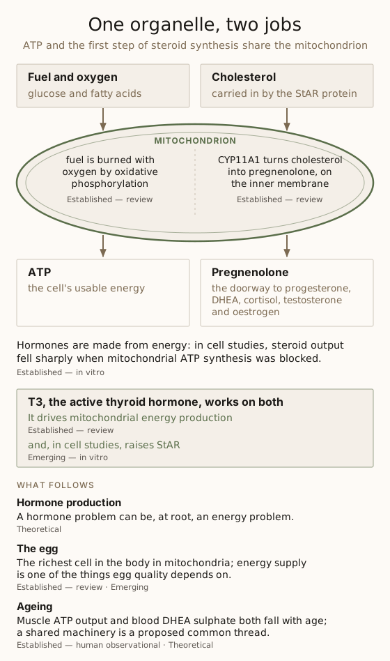
That is the crux, and it is worth reading twice: hormone production starts at the energy organelle. Pregnenolone is the doorway to progesterone, DHEA, cortisol, testosterone, and oestrogen — the whole steroid network. And the door is on the inside.
The convergence, in one line
ATP production and the first step of steroid synthesis both run through the mitochondrion. They are not neighbours. They are housemates.
T3 — the thread running through both
The two jobs are not merely co-located. They share a driver. T3 drives mitochondrial energy production [Established — review],4 and T3 also supports the steroidogenic machinery: in a mouse Leydig cell line, it raised StAR expression about 3.6-fold and progesterone output about fourfold [Emerging — in vitro].6 This is why the thyroid keeps surfacing beneath problems that look unrelated to it: it is a lever on the whole organelle rather than on one output of it [Theoretical].
The overlooked gland takes the same point from the thyroid’s side: how the hormone reaches the cell, and how it is measured.
Why the egg is the sharpest case
If the metabolic-bioenergetic account applies anywhere most sharply, it is fertility, and specifically the oocyte.
The egg is the richest cell in the human body in mitochondria, and it needs them: it depends on them, and on the ATP they make, to become competent for fertilisation and early embryo development [Established — review].7 The embryo inherits essentially all of its mitochondria from the egg [Established — review].7
Mitochondrial function in the oocyte is increasingly understood as a significant contributor to egg quality and developmental competence, and mitochondrial capacity in eggs tends to decline with maternal age [Emerging — review, human and animal].7 That decline is one proposed contributor to the age-related fall in fertility. It has not yet given a usable measurement: in two studies of IVF embryos, the amount of mitochondrial DNA did not differ with the mother’s age and did not predict implantation [Emerging — human observational, null].89
Put plainly: energy supply is one of the things egg quality depends on [Emerging]. The other side is chromosomal, and it changes sharply with age: across 15,169 embryo biopsies, the share of women with no chromosomally normal embryo was 2–6% between 26 and 37, 33% at 42 and 53% at 44 [Established — human observational].10 The energy side is the argument fertility as system consequence makes for the whole organism: that reproduction is a readout of whether the system has energy to spare [Theoretical].
You cannot supplement past an energy problem
The clinical implication is humbling and clarifying at once. If hormone synthesis begins at the energy organelle, then a hormone problem can be, at root, an energy problem — and adjusting hormones downstream does not by itself correct a shortfall upstream [Theoretical].
This reframes the question. Instead of only asking which hormone is low, the metabolic-bioenergetic framework also asks whether the machinery that makes hormones is supplied and running: whether its mitochondria can make ATP by oxidative phosphorylation, and bring cholesterol in to start steroid synthesis. That question brings in thyroid function, energy availability, the substrate itself, the cofactors — among them selenium, zinc and iron, on which thyroid hormone production and conversion depend [Established — review]11 — and the stress and inflammatory load competing for the same supply.
What the machinery needs
| Input | Role | Evidence |
|---|---|---|
| Cholesterol | The substrate — the starting block of every steroid hormone, carried into the mitochondrion by StAR. | Established — review |
| T3 | Pace-setter for energy production; in cell studies, it also raises StAR. | Established — review (energy); Emerging — in vitro (steroid synthesis) |
| Oxygen and fuel | Substrate for oxidative phosphorylation, which makes most of the cell’s ATP. | Established — review |
| Micronutrient cofactors | Including those required for thyroid hormone production and conversion — selenium, zinc, iron. | Established — review |
| Retinoic acid | A vitamin A derivative; in cell studies, it raises StAR and steroid output.12 | Emerging — in vitro |
In a study of 22,748 pregnancies, more than 10,000 IU a day of preformed vitamin A from supplements in early pregnancy went with more birth defects, so this is a mechanism seen in cells, not a reason to take vitamin A [Established — human observational].13
What the supplement trials show
In the Cochrane review of antioxidants for women with subfertility — 63 trials, 7,760 women — the evidence was of such low certainty that the reviewers could not say whether antioxidants raise live births [Established — systematic review, very low-certainty evidence].14 In the MOXI trial, an antioxidant combination given to men with male-factor infertility did not improve sperm concentration, motility, shape or DNA integrity, and live births did not differ: 15% against 24% with placebo [Established — human RCT, null].15
What can actually be changed
This is the point at which mechanism is most often oversold, so I want to be exact about where the leverage actually is.
Nothing reaches inside an organelle and improves it by hand. What can be changed is everything the organelle depends on: the supply of fuel, oxygen and cofactors, thyroid signalling, and the stress and inflammatory load competing for the same supply. Which of those is limiting differs from person to person [Clinical Heuristic], and the metabolic-bioenergetic approach to fertility sets out each of the conditions on which ATP production depends.
The organelle is the explanation. The conditions are the intervention.
What this article does not claim
This is educational, not treatment advice, and nothing here promises conception, a particular hormone result, or a particular IVF outcome. Outcomes are multifactorial and cannot be guaranteed.
Ageing — the parallel decline
Across adult life, two things decline: the capacity of mitochondria to make energy, and steroid hormone output. In 146 healthy adults aged 18 to 89, the ATP production of muscle mitochondria fell with age [Established — human observational];16 and in 1,462 people aged 11 to 89, blood levels of DHEA sulphate, an adrenal steroid made from pregnenolone, peaked in the late teens and early twenties and fell steadily, to about a fifth of the peak after 70 [Established — human observational].17 The metabolic-bioenergetic account offers a parsimonious explanation: both depend, in part, on the same machinery, the mitochondria that make ATP and start steroid synthesis [Theoretical].
Declining mitochondrial function has been proposed as a central thread in ageing more broadly, and it is one of twelve proposed hallmarks of ageing [Established — review].18 That wider mitochondrial theory of ageing remains one explanatory framework among several rather than settled fact [contested]: in mice, 17 of 18 genetic changes to antioxidant enzymes left lifespan unchanged, which casts doubt on its oxidative-damage version [Established — animal, null].19 The narrower observation — that energy production and hormone production share a common and vulnerable machinery — stands on considerably firmer ground.
Key takeaways
- The first, rate-limiting step in making every steroid hormone, the conversion of cholesterol to pregnenolone by the enzyme CYP11A1, takes place inside the mitochondrion, the organelle that makes the cell’s energy [Established — review].
- Thyroid hormone T3 regulates how mitochondria make energy [Established — review] and, in cell studies, raises StAR, the protein that brings cholesterol into the mitochondrion for steroid synthesis [Emerging — in vitro].
- The human egg is the richest cell in the body in mitochondria, and the embryo inherits essentially all of its mitochondria from the egg [Established — review].
- In two studies of IVF embryos, the amount of mitochondrial DNA did not differ with the mother’s age and did not predict implantation [Emerging — human observational, null].
- A hormone problem can be, at root, an energy problem, because the first step of steroid synthesis depends on working mitochondria [Theoretical].
Frequently asked questions
What do mitochondria have to do with hormones?
The first and rate-limiting step in producing every steroid hormone — converting cholesterol to pregnenolone — takes place inside the mitochondrion, performed by the enzyme CYP11A1.1 Energy production and hormone production therefore share the same location and several of the same drivers, principally the active thyroid hormone T3.
Can you treat mitochondria?
Not as a target in its own right. What can be influenced are the conditions under which cells produce energy: thyroid signalling, nutrient and cofactor availability, oxidative and inflammatory load, sleep, and the competing demands on the same supply.
Do mitochondria affect egg quality?
The oocyte is the richest cell in the body in mitochondria, and the embryo inherits essentially all of its mitochondria from it. Research increasingly links oocyte mitochondrial function to egg quality and developmental competence.7 That link is an emerging research direction rather than a settled determinant: so far, the amount of mitochondrial DNA in an embryo has not predicted whether it implants.89
Is the mitochondrial theory of ageing established?
No. It is one explanatory framework among several and remains contested as an account of primary cause.19 The narrower point — that energy production and steroid hormone production share common machinery that becomes less efficient with age — rests on firmer evidence.16
Where does thyroid function fit in?
T3 sets the pace of mitochondrial energy production4 and, in cell studies, supports the first step of steroid synthesis.6 The overlooked gland explains how thyroid hormone is made, converted and measured.
References
-
Miller WL, Auchus RJ. The molecular biology, biochemistry, and physiology of human steroidogenesis and its disorders. Endocr Rev. 2011;32(1):81–151. doi:10.1210/er.2010-0013. PMID: 21051590. [Established — review] ↩︎↩︎↩︎
-
Allen JA, Shankara T, Janus P, Buck S, Diemer T, Hales KH, et al. Energized, polarized, and actively respiring mitochondria are required for acute Leydig cell steroidogenesis. Endocrinology. 2006;147(8):3924–3935. doi:10.1210/en.2005-1204. PMID: 16690799. [Established — in vitro] ↩︎
-
Nolfi-Donegan D, Braganza A, Shiva S. Mitochondrial electron transport chain: oxidative phosphorylation, oxidant production, and methods of measurement. Redox Biol. 2020;37:101674. doi:10.1016/j.redox.2020.101674. PMID: 32811789. [Established — review] ↩︎
-
Weitzel JM, Iwen KA. Coordination of mitochondrial biogenesis by thyroid hormone. Mol Cell Endocrinol. 2011;342(1–2):1–7. doi:10.1016/j.mce.2011.05.009. PMID: 21664416. [Established — review] ↩︎↩︎↩︎
-
Miller WL. Steroid hormone synthesis in mitochondria. Mol Cell Endocrinol. 2013;379(1–2):62–73. doi:10.1016/j.mce.2013.04.014. PMID: 23628605. [Established — review] ↩︎
-
Manna PR, Tena-Sempere M, Huhtaniemi IT. Molecular mechanisms of thyroid hormone-stimulated steroidogenesis in mouse Leydig tumor cells: involvement of the steroidogenic acute regulatory (StAR) protein. J Biol Chem. 1999;274(9):5909–5918. doi:10.1074/jbc.274.9.5909. PMID: 10026215. [Emerging — in vitro] ↩︎↩︎
-
May-Panloup P, Boucret L, Chao de la Barca JM, Desquiret-Dumas V, Ferré-L’Hotellier V, Morinière C, et al. Ovarian ageing: the role of mitochondria in oocytes and follicles. Hum Reprod Update. 2016;22(6):725–743. doi:10.1093/humupd/dmw028. PMID: 27562289. [Established — review] ↩︎↩︎↩︎↩︎
-
Victor AR, Brake AJ, Tyndall JC, Griffin DK, Zouves CG, Barnes FL, et al. Accurate quantitation of mitochondrial DNA reveals uniform levels in human blastocysts irrespective of ploidy, age, or implantation potential. Fertil Steril. 2017;107(1):34–42.e3. doi:10.1016/j.fertnstert.2016.09.028. PMID: 27793366. [Emerging — human observational, null] ↩︎↩︎
-
Treff NR, Zhan Y, Tao X, Olcha M, Han M, Rajchel J, et al. Levels of trophectoderm mitochondrial DNA do not predict the reproductive potential of sibling embryos. Hum Reprod. 2017;32(4):954–962. doi:10.1093/humrep/dex034. PMID: 28333210. [Emerging — human observational, null] ↩︎↩︎
-
Franasiak JM, Forman EJ, Hong KH, Werner MD, Upham KM, Treff NR, et al. The nature of aneuploidy with increasing age of the female partner: a review of 15,169 consecutive trophectoderm biopsies evaluated with comprehensive chromosomal screening. Fertil Steril. 2014;101(3):656–663.e1. doi:10.1016/j.fertnstert.2013.11.004. PMID: 24355045. [Established — human observational] ↩︎
-
Zimmermann MB, Köhrle J. The impact of iron and selenium deficiencies on iodine and thyroid metabolism: biochemistry and relevance to public health. Thyroid. 2002;12(10):867–878. doi:10.1089/105072502761016494. PMID: 12487769. [Established — review] ↩︎
-
Manna PR, Stetson CL, Daugherty C, Shimizu I, Syapin PJ, Garrel G, et al. Up-regulation of steroid biosynthesis by retinoid signaling: implications for aging. Mech Ageing Dev. 2015;150:74–82. doi:10.1016/j.mad.2015.08.007. PMID: 26303142. [Emerging — in vitro] ↩︎
-
Rothman KJ, Moore LL, Singer MR, Nguyen US, Mannino S, Milunsky A. Teratogenicity of high vitamin A intake. N Engl J Med. 1995;333(21):1369–1373. doi:10.1056/NEJM199511233332101. PMID: 7477116. [Established — human observational] ↩︎
-
Showell MG, Mackenzie-Proctor R, Jordan V, Hart RJ. Antioxidants for female subfertility. Cochrane Database Syst Rev. 2020;8(8):CD007807. doi:10.1002/14651858.CD007807.pub4. PMID: 32851663. [Established — systematic review, very low-certainty evidence] ↩︎
-
Steiner AZ, Hansen KR, Barnhart KT, Cedars MI, Legro RS, Diamond MP, et al. The effect of antioxidants on male factor infertility: the Males, Antioxidants, and Infertility (MOXI) randomized clinical trial. Fertil Steril. 2020;113(3):552–560.e3. doi:10.1016/j.fertnstert.2019.11.008. PMID: 32111479. [Established — human RCT, null] ↩︎
-
Short KR, Bigelow ML, Kahl J, Singh R, Coenen-Schimke J, Raghavakaimal S, et al. Decline in skeletal muscle mitochondrial function with aging in humans. Proc Natl Acad Sci U S A. 2005;102(15):5618–5623. doi:10.1073/pnas.0501559102. PMID: 15800038. [Established — human observational] ↩︎↩︎
-
Orentreich N, Brind JL, Rizer RL, Vogelman JH. Age changes and sex differences in serum dehydroepiandrosterone sulfate concentrations throughout adulthood. J Clin Endocrinol Metab. 1984;59(3):551–555. doi:10.1210/jcem-59-3-551. PMID: 6235241. [Established — human observational] ↩︎
-
López-Otín C, Blasco MA, Partridge L, Serrano M, Kroemer G. Hallmarks of aging: an expanding universe. Cell. 2023;186(2):243–278. doi:10.1016/j.cell.2022.11.001. PMID: 36599349. [Established — review] ↩︎
-
Pérez VI, Bokov A, Van Remmen H, Mele J, Ran Q, Ikeno Y, et al. Is the oxidative stress theory of aging dead? Biochim Biophys Acta. 2009;1790(10):1005–1014. doi:10.1016/j.bbagen.2009.06.003. PMID: 19524016. [Established — animal, null] ↩︎↩︎
Related reading
- The metabolic-bioenergetic approach to fertility — the framework this mechanism sits inside
- The overlooked gland: thyroid hormone and fertility — the shared driver, from the other direction
- Fertility as system consequence — the same argument, for the whole organism