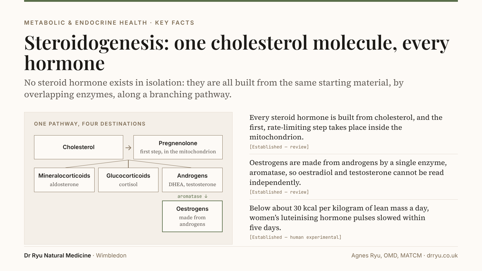
A patient is told their testosterone is “high”, or their progesterone “low”, and a conclusion is drawn from that single figure. The trouble is that no steroid hormone exists in isolation. They are all built from the same starting material, by overlapping enzymes, along a branching pathway where the output of one step is the raw material of the next. Change one part and you change others.
A number only means something once you can see where it sits in the network. This article maps the network.
One origin — everything begins as cholesterol
Here is the fact that surprises almost everyone: cortisol, progesterone, DHEA, testosterone, oestrogen, and aldosterone are all built from cholesterol. There is no separate raw material for “sex” hormones and “stress” hormones. They share a single source [Established — review].1
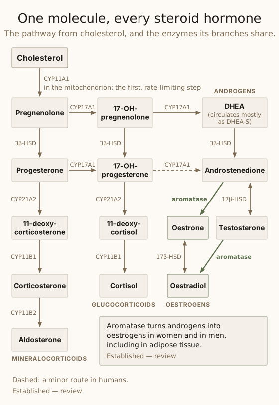
The first and rate-limiting step — cholesterol converted to pregnenolone by the enzyme CYP11A1 — happens inside the mitochondrion, the cell’s energy compartment [Established — review].1 That detail is not incidental. It is the physical bridge between hormone production and energy production, and it has its own article: The Mitochondrion — Where Energy Meets Hormones.
Where the cholesterol comes from
Steroid-making cells take cholesterol from circulating lipoproteins and make some of their own [Established — review].1 Supply does not become the limit at the levels treatment reaches: in a year-long randomised trial of 901 people whose LDL cholesterol was lowered to very low levels, gonadal hormones did not change and adrenal function held, including in those whose LDL fell below 0.4 mmol/L [Emerging — human RCT].2
One pathway, four destinations
From pregnenolone, the pathway branches into four families, all drawing on a shared toolkit of enzymes:
- Mineralocorticoids (aldosterone) — fluid and blood-pressure regulation
- Glucocorticoids (cortisol) — the stress and metabolic axis
- Androgens (DHEA, testosterone) — the building hormones
- Oestrogens (oestradiol, oestrone, oestriol) — made from androgens
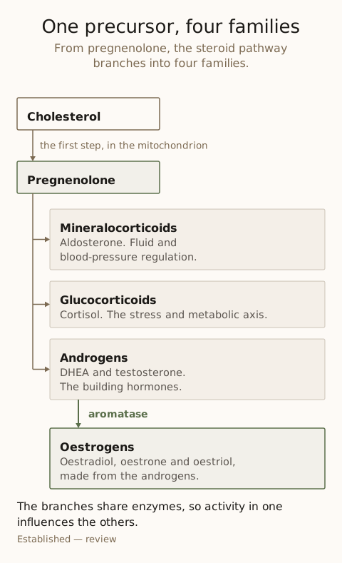
Because the enzymes are shared, activity in one branch influences the others [Established — review].1 This is a genuinely interconnected network, not a set of separate taps.
The hormones, and what they actually do
Progesterone
More than a pregnancy hormone. Progesterone is both a precursor in the pathway and an active hormone in its own right. It dominates the second half of the menstrual cycle, when it transforms the uterine lining for implantation [Established — review],3 and it acts in the brain as a calming neurosteroid through its metabolite allopregnanolone, which strengthens the action of GABA, the brain’s main inhibitory signal [Established — review].4 It is made in quantity only after ovulation, which is why a mid-luteal progesterone is the standard check that ovulation has occurred [Established — clinical guideline],5 and why progesterone is best read in the context of the cycle rather than as a stray number.
DHEA
Made largely by the adrenal glands. DHEA and its sulfated form, DHEA-S, which circulates at a higher concentration than any other androgen, constitute a precursor reservoir the body draws on to make both androgens and oestrogens in peripheral tissues [Established — review]6 — particularly relevant after menopause, when ovarian production steps back. DHEA-S declines steadily from its peak in early adulthood [Established — human observational].7
Cortisol
The principal glucocorticoid, produced continuously and in substantial amounts to run the stress and metabolic axis. Cortisol is broadly catabolic: it raises glucose production, mobilises amino acids from muscle and stimulates fat breakdown [Established — review].8 In short bursts that is adaptive. Sustained demand is where problems accumulate.
What turns the pathway down
The pathway runs on energy. Its first step takes place inside the mitochondrion, and in a mouse Leydig cell line steroid production fell sharply when the mitochondria could not make energy [Established — in vitro];9 the argument is set out in The Mitochondrion — Where Energy Meets Hormones. Three common states turn the pathway down. Energy deficiency disrupts the signal that drives the ovary: when young women’s energy availability was held below about 30 kcal per kilogram of lean mass a day for five days, their pulses of luteinising hormone became less frequent [Established — human experimental].10 Sustained stress acts through cortisol on the hypothalamus, the pituitary and the gonads, and turns the reproductive axis down [Established — review].8 And an underactive thyroid alters the sex steroids and the protein that carries them in the blood, and goes with infrequent periods and reduced fertility [Established — review];11 see The Overlooked Gland.
Testosterone
An androgen — and not a male-only hormone. Women make and need testosterone too: in women it circulates at higher concentrations than oestradiol, and it bears on sexual desire, muscle and bone [Established — review].12 It is a building (anabolic) hormone in both sexes. Crucially, testosterone is built through the pathway rather than summoned, which is why raising it without reading the upstream network can miss the cause [Clinical Heuristic].
Oestrogen — and the aromatase surprise
Oestrogens are not made from a separate precursor. They are made from androgens. Testosterone is converted to oestradiol, and androstenedione to oestrone, by a single enzyme: aromatase [Established — review].13 This is why oestrogen and testosterone can never be read independently. They are two ends of the same reaction.
Aromatisation — the same enzyme in men and women
Aromatase converts androgens into oestrogens, and it operates in both sexes. Men make oestrogen from their testosterone; women make testosterone, some of which becomes oestrogen.
The enzyme is active not only in the gonads but in adipose tissue, bone and brain; after menopause, and in men, much of the body’s oestradiol is made in these tissues [Established — review].13 That is one reason body composition influences the androgen-to-oestrogen ratio: in men, the conversion of testosterone to oestradiol rises in proportion to excess weight [Established — human observational].14
This single fact reframes a great deal. A high-oestrogen picture in a man, or a shifting androgen-to-oestrogen ratio through perimenopause, often makes sense only once you look at the androgens it came from and the aromatase converting them [Clinical Heuristic].
The adrenal glands — the second hormone factory
The adrenal glands run the same steroid pathway as the gonads, each zone of the adrenal cortex with its own enzymes: the outer zones make aldosterone and cortisol, the inner zone DHEA and DHEA-S [Established — review].15 Through DHEA, the adrenals contribute to the body’s androgen and oestrogen supply [Established — review].6
This matters most when ovarian production winds down: after menopause, peripheral conversion of adrenal DHEA becomes a meaningful source of sex hormones [Established — review].613 The adrenal network is not separate from the reproductive one. They share the same machinery and the same raw materials.
Building versus breaking down
One useful way to read the steroid network is as two opposing demands.
| Direction | Principal hormones | |
|---|---|---|
| Catabolic | Breaking down — mobilising energy from tissue | Cortisol |
| Anabolic | Building up — repair and maintenance | DHEA, testosterone |
The two are meant to alternate: mobilisation under demand, repair in recovery [Theoretical]. Severe illness shows the shift at its extreme: in 30 patients with septic shock, DHEA-S on admission was less than a third of the level in matched controls, while cortisol stayed raised throughout [Established — human observational].16 Whether ordinary sustained stress or under-recovery does the same is less certain; in one study of 81 working adults, those who reported stress at work had DHEA-S levels 23% lower, which is why the ratio of cortisol to DHEA-S is studied as one window onto the shift [Emerging — human observational].17
Why this means reading the whole network
Now the practical payoff. Because the hormones share an origin, share enzymes, and convert into one another, a single result is rarely interpretable alone [Clinical Heuristic].
- A “high” testosterone may reflect production, or poor conversion onward, or reduced aromatisation.
- A “low” oestrogen cannot be understood without the androgens it is made from.
- A progesterone reading is only meaningful in the context of ovulation and cycle timing.
- Cortisol demand colours the entire system, not only the stress axis.
This is also why some tests report metabolites rather than levels alone — how hormones are being broken down and routed, not simply how much is present at one moment [Established — review].18 What such a profile reports is set out on the steroid hormone testing page.
Key takeaways
- Every steroid hormone — cortisol, progesterone, DHEA, testosterone, the oestrogens and aldosterone — is built from cholesterol, and the first, rate-limiting step takes place inside the mitochondrion [Established — review].
- Oestrogens are made from androgens by a single enzyme, aromatase, so oestradiol and testosterone cannot be read independently of each other [Established — review].
- Aromatase is active in adipose tissue as well as the gonads, and in men the conversion of testosterone to oestradiol rises in proportion to excess weight [Established — human observational].
- Energy deficiency turns down the signal that drives the ovary: below about 30 kcal per kilogram of lean mass a day, women’s pulses of luteinising hormone became less frequent within five days [Established — human experimental].
- Because the steroid hormones share a precursor and enzymes and convert into one another, a single hormone result read on its own misleads more often than it informs [Clinical Heuristic].
Frequently asked questions
What is steroidogenesis?
It is the process by which the body builds steroid hormones — progesterone, DHEA, cortisol, testosterone, oestrogen, and aldosterone — from a single starting material, cholesterol, along a branching pathway of shared enzymes. Because the enzymes are shared, the hormones form a connected network rather than independent outputs.
Is oestrogen really made from testosterone?
Yes. The enzyme aromatase converts testosterone to oestradiol and androstenedione to oestrone. This happens in both sexes, and not only in the gonads — adipose tissue is an active site of aromatase activity.
Why does cholesterol matter for hormones?
It is the substrate from which every steroid hormone, including progesterone, cortisol, testosterone and oestrogen, is built.
References
-
Miller WL, Auchus RJ. The molecular biology, biochemistry, and physiology of human steroidogenesis and its disorders. Endocr Rev. 2011;32(1):81–151. doi:10.1210/er.2010-0013. PMID: 21051590. [Established — review] ↩︎↩︎↩︎↩︎
-
Blom DJ, Djedjos CS, Monsalvo ML, Bridges I, Wasserman SM, Scott R, et al. Effects of evolocumab on vitamin E and steroid hormone levels: results from the 52-week, phase 3, double-blind, randomized, placebo-controlled DESCARTES study. Circ Res. 2015;117(8):731–741. doi:10.1161/CIRCRESAHA.115.307071. PMID: 26228031. [Emerging — human RCT] ↩︎
-
Gellersen B, Brosens JJ. Cyclic decidualization of the human endometrium in reproductive health and failure. Endocr Rev. 2014;35(6):851–905. doi:10.1210/er.2014-1045. PMID: 25141152. [Established — review] ↩︎
-
Paul SM, Purdy RH. Neuroactive steroids. FASEB J. 1992;6(6):2311–2322. PMID: 1347506. [Established — review] ↩︎
-
National Institute for Health and Care Excellence. Fertility problems: assessment and treatment (NG257). Published 31 March 2026. Recommendation 1.18.7. https://www.nice.org.uk/guidance/ng257 [Established — clinical guideline] ↩︎
-
Burger HG. Androgen production in women. Fertil Steril. 2002;77(Suppl 4):S3–S5. doi:10.1016/s0015-0282(02)02985-0. PMID: 12007895. [Established — review] ↩︎↩︎↩︎
-
Orentreich N, Brind JL, Rizer RL, Vogelman JH. Age changes and sex differences in serum dehydroepiandrosterone sulfate concentrations throughout adulthood. J Clin Endocrinol Metab. 1984;59(3):551–555. doi:10.1210/jcem-59-3-551. PMID: 6235241. [Established — human observational] ↩︎
-
Whirledge S, Cidlowski JA. Glucocorticoids, stress, and fertility. Minerva Endocrinol. 2010;35(2):109–125. PMID: 20595939. [Established — review] ↩︎↩︎
-
Allen JA, Shankara T, Janus P, Buck S, Diemer T, Hales KH, et al. Energized, polarized, and actively respiring mitochondria are required for acute Leydig cell steroidogenesis. Endocrinology. 2006;147(8):3924–3935. doi:10.1210/en.2005-1204. PMID: 16690799. [Established — in vitro] ↩︎
-
Loucks AB, Thuma JR. Luteinizing hormone pulsatility is disrupted at a threshold of energy availability in regularly menstruating women. J Clin Endocrinol Metab. 2003;88(1):297–311. doi:10.1210/jc.2002-020369. PMID: 12519869. [Established — human experimental] ↩︎
-
Krassas GE, Poppe K, Glinoer D. Thyroid function and human reproductive health. Endocr Rev. 2010;31(5):702–755. doi:10.1210/er.2009-0041. PMID: 20573783. [Established — review] ↩︎
-
Davis SR, Wahlin-Jacobsen S. Testosterone in women — the clinical significance. Lancet Diabetes Endocrinol. 2015;3(12):980–992. doi:10.1016/S2213-8587(15)00284-3. PMID: 26358173. [Established — review] ↩︎
-
Simpson ER. Sources of estrogen and their importance. J Steroid Biochem Mol Biol. 2003;86(3–5):225–230. doi:10.1016/s0960-0760(03)00360-1. PMID: 14623515. [Established — review] ↩︎↩︎↩︎
-
Schneider G, Kirschner MA, Berkowitz R, Ertel NH. Increased estrogen production in obese men. J Clin Endocrinol Metab. 1979;48(4):633–638. doi:10.1210/jcem-48-4-633. PMID: 429508. [Established — human observational] ↩︎
-
Rainey WE, Nakamura Y. Regulation of the adrenal androgen biosynthesis. J Steroid Biochem Mol Biol. 2008;108(3–5):281–286. doi:10.1016/j.jsbmb.2007.09.015. PMID: 17945481. [Established — review] ↩︎
-
Beishuizen A, Thijs LG, Vermes I. Decreased levels of dehydroepiandrosterone sulphate in severe critical illness: a sign of exhausted adrenal reserve? Crit Care. 2002;6(5):434–438. doi:10.1186/cc1530. PMID: 12398784. [Established — human observational] ↩︎
-
Lennartsson AK, Theorell T, Rockwood AL, Kushnir MM, Jonsdottir IH. Perceived stress at work is associated with lower levels of DHEA-S. PLoS One. 2013;8(8):e72460. doi:10.1371/journal.pone.0072460. PMID: 24015247. [Emerging — human observational] ↩︎
-
Storbeck KH, Schiffer L, Baranowski ES, Chortis V, Prete A, Barnard L, et al. Steroid metabolome analysis in disorders of adrenal steroid biosynthesis and metabolism. Endocr Rev. 2019;40(6):1605–1625. doi:10.1210/er.2018-00262. PMID: 31294783. [Established — review] ↩︎
Related reading
- The Mitochondrion — Where Energy Meets Hormones — where the first step physically happens
- The Overlooked Gland — Thyroid Hormone and Fertility — the thyroid’s pace-setting role across the network
- Fertility as System Consequence — why stress and energy reach reproduction through the whole system
- Metabolic Fertility Testing — when a wider look than a standard panel is worth taking